Blog

  • Uncovering the spectrum of adult zebrafish neural stem cell cycle regulators

    Uncovering the spectrum of adult zebrafish neural stem cell cycle regulators

    Aurelien Caron, Lidia Trzuskot, Benjamin W Lindsey

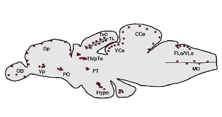
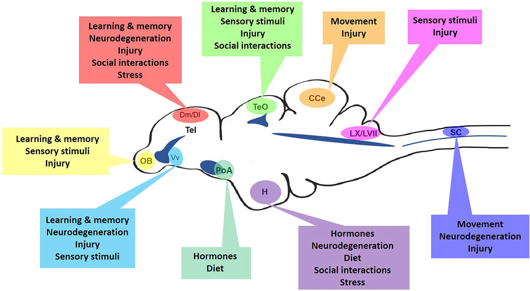
    Adult neural stem and progenitor cells (aNSPCs) persist lifelong in teleost models in diverse stem cell niches of the brain and spinal cord. Fish maintain developmental stem cell populations throughout life, including both neuro-epithelial cells (NECs) and radial-glial cells (RGCs). Within stem cell domains of the brain, RGCs persist in a cycling or quiescent state, whereas NECs continuously divide. Heterogeneous populations of RGCs also sit adjacent the central canal of the spinal cord, showing infrequent proliferative activity under homeostasis. With the rise of the zebrafish (Danio rerio) model to study adult neurogenesis and neuroregeneration in the central nervous system (CNS), it has become evident that aNSPC proliferation is regulated by a wealth of stimuli that may be coupled with biological function. Growing evidence suggests that aNSPCs are sensitive to environmental cues, social interactions, nutrient availability, and neurotrauma for example, and that distinct stem and progenitor cell populations alter their cell cycle activity accordingly. Such stimuli appear to act as triggers to either turn on normally dormant aNSPCs or modulate constitutive rates of niche-specific cell cycle behaviour. Defining the various forms of stimuli that influence RGC and NEC proliferation, and identifying the molecular regulators responsible, will strengthen our understanding of the connection between aNSPC activity and their biological significance. In this review, we aim to bring together the current state of knowledge on aNSPCs from studies investigating the zebrafish CNS, while highlighting emerging cell cycle regulators and outstanding questions that will help to advance this fascinating field of stem cell biology.


    READ FULL PUBLICATION

    Caron et al., 2022 (960 KB)

  • TEM, SEM, and STEM‑based immuno‑CLEM workflows offer complementary advantages

    TEM, SEM, and STEM‑based immuno‑CLEM workflows offer complementary advantages

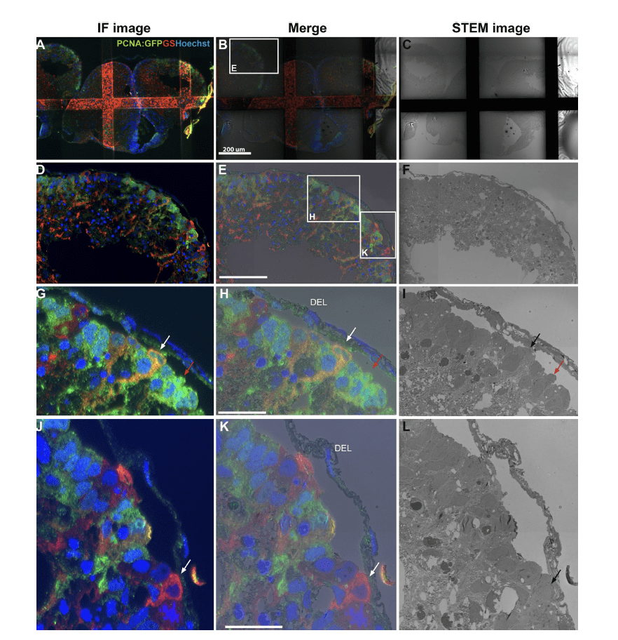
    [vc_row][vc_column][ultimate_spacer height=”35″ height_on_tabs=”25″ height_on_tabs_portrait=”25″ height_on_mob_landscape=”25″ height_on_mob=”25″][vc_custom_heading text=”SCIENTIFIC REPORTS” font_container=”tag:p|text_align:left|line_height:1.3em” google_fonts=”font_family:Muli%3A300%2C300italic%2Cregular%2Citalic|font_style:400%20regular%3A400%3Anormal” css_animation=”slideInLeft”][ultimate_spacer height=”10″ height_on_tabs=”10″ height_on_tabs_portrait=”10″ height_on_mob_landscape=”10″ height_on_mob=”10″][vc_custom_heading text=”TEM, SEM, and STEM‑based immuno‑CLEM workflows offer complementary advantages” font_container=”tag:h3|text_align:left|line_height:1.3em” google_fonts=”font_family:Muli%3A300%2C300italic%2Cregular%2Citalic|font_style:400%20regular%3A400%3Anormal” css_animation=”slideInLeft”][ultimate_spacer height=”10″ height_on_tabs=”10″ height_on_tabs_portrait=”10″ height_on_mob_landscape=”10″ height_on_mob=”10″][vc_column_text]Viola Oorschot, Benjamin W Lindsey, Jan Kaslin & Georg Ramm[/vc_column_text][ultimate_spacer height=”25″ height_on_tabs=”25″ height_on_tabs_portrait=”25″ height_on_mob_landscape=”25″ height_on_mob=”25″][vc_column_text]Identifying endogenous tissue stem cells remains a key challenge in developmental and regenerative biology. To distinguish and molecularly characterise stem cell populations in large heterogeneous tissues, the combination of cytochemical cell markers with ultrastructural morphology is highly beneficial. Here, we realise this through workflows of multi-resolution immuno-correlative light and electron microscopy (iCLEM) methodologies. Taking advantage of the antigenicity preservation of the Tokuyasu technique, we have established robust protocols and workflows and provide a sideby-side comparison of iCLEM used in combination with scanning EM (SEM), scanning TEM (STEM), or transmission EM (TEM). Evaluation of the applications and advantages of each method highlights their practicality for the identification, quantification, and characterization of heterogeneous cell populations in small organisms, organs, or tissues in healthy and diseased states. The iCLEM techniques are broadly applicable and can use either genetically encoded or cytochemical markers on plant, animal and human tissues. We demonstrate how these protocols are particularly suited for investigating neural stem and progenitor cell populations of the vertebrate nervous system.[/vc_column_text][ultimate_spacer height=”25″ height_on_tabs=”25″ height_on_tabs_portrait=”25″ height_on_mob_landscape=”25″ height_on_mob=”25″][vc_column_text]READ FULL PUBLICATION

    Oorschot et al., 2021 (4.4 MB)

    [/vc_column_text][ultimate_spacer height=”25″ height_on_tabs=”25″ height_on_tabs_portrait=”25″ height_on_mob_landscape=”25″ height_on_mob=”25″][vc_btn title=”back to publications” color=”orange” i_icon_fontawesome=”fa fa-bookmark-o” add_icon=”true” link=”url:http%3A%2F%2Flindseylab.ca%2Fpublications%2F|title:PUBLICATIONS%202||”][ultimate_spacer height=”25″ height_on_tabs=”25″ height_on_tabs_portrait=”25″ height_on_mob_landscape=”25″ height_on_mob=”25″][/vc_column][/vc_row]

  • Spatiotemporal Transition in the Role of Synaptic Inhibition to the Tail Beat Rhythm of Developing Larval Zebrafish

    Spatiotemporal Transition in the Role of Synaptic Inhibition to the Tail Beat Rhythm of Developing Larval Zebrafish

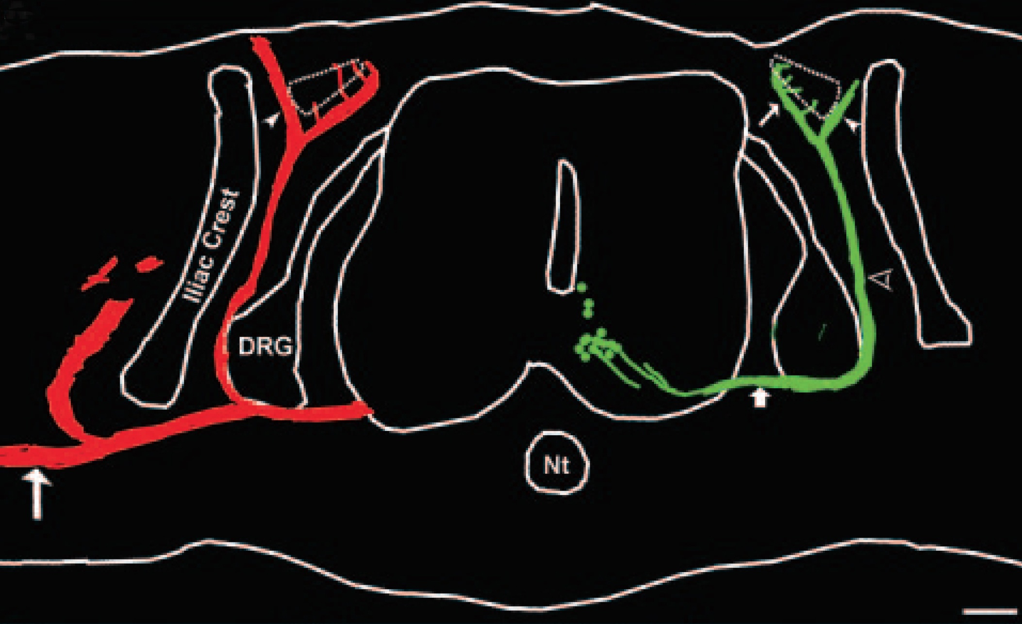
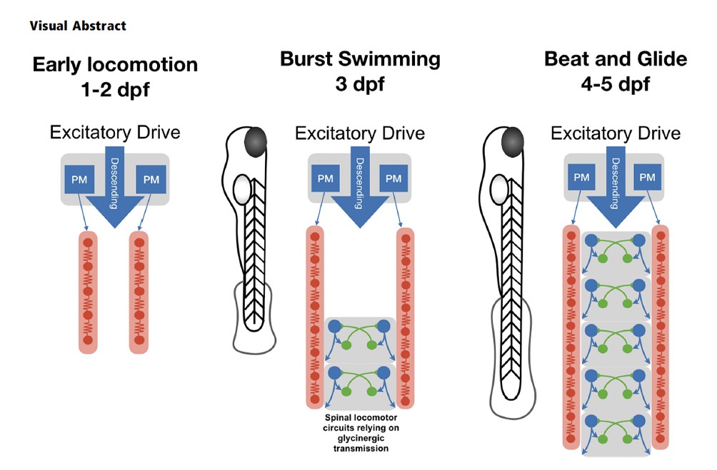
    [vc_row][vc_column][ultimate_spacer height=”35″ height_on_tabs=”25″ height_on_tabs_portrait=”25″ height_on_mob_landscape=”25″ height_on_mob=”25″][vc_custom_heading text=”ENEURO” font_container=”tag:p|text_align:left|line_height:1.3em” google_fonts=”font_family:Muli%3A300%2C300italic%2Cregular%2Citalic|font_style:400%20regular%3A400%3Anormal” css_animation=”slideInLeft”][ultimate_spacer height=”10″ height_on_tabs=”10″ height_on_tabs_portrait=”10″ height_on_mob_landscape=”10″ height_on_mob=”10″][vc_custom_heading text=”Spatiotemporal Transition in the Role of Synaptic Inhibition to the Tail Beat Rhythm of Developing Larval Zebrafish” font_container=”tag:h3|text_align:left|line_height:1.3em” google_fonts=”font_family:Muli%3A300%2C300italic%2Cregular%2Citalic|font_style:400%20regular%3A400%3Anormal” css_animation=”slideInLeft”][ultimate_spacer height=”10″ height_on_tabs=”10″ height_on_tabs_portrait=”10″ height_on_mob_landscape=”10″ height_on_mob=”10″][vc_column_text]Yann Roussel, Melissa Paradis, Stephanie F. Gaudreau, Benjamin W Lindsey, and Tuan V. Bui[/vc_column_text][ultimate_spacer height=”25″ height_on_tabs=”25″ height_on_tabs_portrait=”25″ height_on_mob_landscape=”25″ height_on_mob=”25″][vc_column_text]Significant maturation of swimming in zebrafish (Danio rerio) occurs within the first few days of life when fish transition from coiling movements to burst swimming and then to beat-and-glide swimming. This maturation occurs against a backdrop of numerous developmental changes – neurogenesis, a transition from predominantly electrical to chemical-based neurotransmission, and refinement of intrinsic properties. There is evidence that spinal locomotor circuits undergo fundamental changes as the zebrafish transitions from burst to beat-and-glide swimming. Our electrophysiological recordings confirm that the operation of spinal locomotor circuits becomes increasingly reliant on glycinergic neurotransmission for rhythmogenesis governing the rhythm of tail beats. This transition occurred at the same time that we observed a change in rhythmicity of synaptic inhibition to spinal motoneurons (MNs). When we examined whether the transition from weakly to strongly glycinergic dependent rhythmogenesis occurred at a uniform pace across the length of the spinal cord, we found that this transition occurred earlier at caudal segments than at rostral segments of the spinal cord. Furthermore, while this rhythmogenic transition occurred when fish transition from burst swimming to beat-and-glide swimming, these two transitions were not interdependent. These results suggest that there is a developmental transition in the operation of spinal locomotor circuits that is gradually set in place in the spinal cord in a caudo-rostral temporal sequence.[/vc_column_text][ultimate_spacer height=”25″ height_on_tabs=”25″ height_on_tabs_portrait=”25″ height_on_mob_landscape=”25″ height_on_mob=”25″][vc_column_text]READ FULL PUBLICATION

    Roussel et al., 2020 (4.3 MB)

    [/vc_column_text][ultimate_spacer height=”25″ height_on_tabs=”25″ height_on_tabs_portrait=”25″ height_on_mob_landscape=”25″ height_on_mob=”25″][vc_btn title=”back to publications” color=”orange” i_icon_fontawesome=”fa fa-bookmark-o” add_icon=”true” link=”url:http%3A%2F%2Flindseylab.ca%2Fpublications%2F|title:PUBLICATIONS%202||”][ultimate_spacer height=”25″ height_on_tabs=”25″ height_on_tabs_portrait=”25″ height_on_mob_landscape=”25″ height_on_mob=”25″][/vc_column][/vc_row]
  • Midbrain tectal stem cells display diverse regenerative capacities in zebrafish

    Midbrain tectal stem cells display diverse regenerative capacities in zebrafish

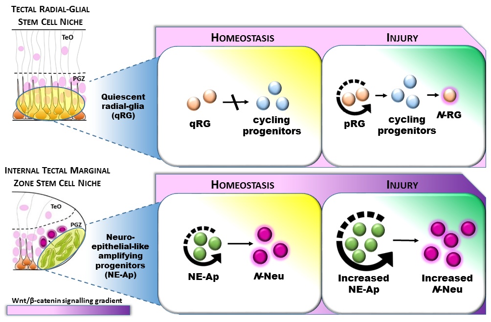
    [vc_row][vc_column][ultimate_spacer height=”35″ height_on_tabs=”25″ height_on_tabs_portrait=”25″ height_on_mob_landscape=”25″ height_on_mob=”25″][vc_custom_heading text=”SCIENTIFIC REPORTS” font_container=”tag:p|text_align:left|line_height:1.3em” google_fonts=”font_family:Muli%3A300%2C300italic%2Cregular%2Citalic|font_style:400%20regular%3A400%3Anormal” css_animation=”slideInLeft”][ultimate_spacer height=”10″ height_on_tabs=”10″ height_on_tabs_portrait=”10″ height_on_mob_landscape=”10″ height_on_mob=”10″][vc_custom_heading text=”Midbrain tectal stem cells display diverse regenerative capacities in zebrafish” font_container=”tag:h3|text_align:left|line_height:1.3em” google_fonts=”font_family:Muli%3A300%2C300italic%2Cregular%2Citalic|font_style:400%20regular%3A400%3Anormal” css_animation=”slideInLeft”][ultimate_spacer height=”10″ height_on_tabs=”10″ height_on_tabs_portrait=”10″ height_on_mob_landscape=”10″ height_on_mob=”10″][vc_column_text]Benjamin W Lindsey, Georgia E. Aitken, Jean K. Tang, Mitra Khabooshan, Alon M. Douek, Celia Vandestadt, and Jan Kaslin[/vc_column_text][ultimate_spacer height=”25″ height_on_tabs=”25″ height_on_tabs_portrait=”25″ height_on_mob_landscape=”25″ height_on_mob=”25″][vc_column_text]How diverse adult stem and progenitor populations regenerate tissue following damage to the brain is poorly understood. In highly regenerative vertebrates, such as zebrafish, radial-glia (RG) and neuroepithelial-like (NE) stem/progenitor cells contribute to neuronal repair after injury. However, not all RG act as neural stem/progenitor cells during homeostasis in the zebrafish brain, questioning the role of quiescent RG (qRG) post-injury. To understand the function of qRG during regeneration, we performed a stab lesion in the adult midbrain tectum to target a population of homeostatic qRG, and investigated their proliferative behaviour, differentiation potential, and Wnt/β-catenin signalling. EdU-labelling showed a small number of proliferating qRG after injury (pRG) but that progeny are restricted to RG. However, injury promoted proliferation of NE progenitors in the internal tectal marginal zone (TMZi) resulting in amplified regenerative neurogenesis. Increased Wnt/β-catenin signalling was detected in TMZi after injury whereas homeostatic levels of Wnt/β-catenin signalling persisted in qRG/pRG. Attenuation of Wnt signalling suggested that the proliferative response post-injury was Wnt/β-catenin-independent. Our results demonstrate that qRG in the tectum have restricted capability in neuronal repair, highlighting that RG have diverse functions in the zebrafish brain. Furthermore, these findings suggest that endogenous stem cell compartments compensate lost tissue by amplifying homeostatic growth.[/vc_column_text][ultimate_spacer height=”25″ height_on_tabs=”25″ height_on_tabs_portrait=”25″ height_on_mob_landscape=”25″ height_on_mob=”25″][vc_column_text]READ FULL PUBLICATION

    Lindsey et al., 2019 (4.1 MB)

    Lindsey et al., 2019 supplementary (3.3 MB)[/vc_column_text][ultimate_spacer height=”25″ height_on_tabs=”25″ height_on_tabs_portrait=”25″ height_on_mob_landscape=”25″ height_on_mob=”25″][vc_btn title=”back to publications” color=”orange” i_icon_fontawesome=”fa fa-bookmark-o” add_icon=”true” link=”url:http%3A%2F%2Flindseylab.ca%2Fpublications%2F|title:PUBLICATIONS%202||”][ultimate_spacer height=”25″ height_on_tabs=”25″ height_on_tabs_portrait=”25″ height_on_mob_landscape=”25″ height_on_mob=”25″][/vc_column][/vc_row]
  • Dr. Lindsey and Dr. Atukorallaya awarded funding for first Zebrafish Facility at University of Manitoba

    [vc_row][vc_column][ultimate_spacer height=”35″ height_on_tabs=”25″ height_on_tabs_portrait=”25″ height_on_mob_landscape=”25″ height_on_mob=”25″][vc_custom_heading text=”JANUARY 2019″ font_container=”tag:p|text_align:left|line_height:1.3em” google_fonts=”font_family:Muli%3A300%2C300italic%2Cregular%2Citalic|font_style:400%20regular%3A400%3Anormal” css_animation=”slideInLeft”][ultimate_spacer height=”10″ height_on_tabs=”10″ height_on_tabs_portrait=”10″ height_on_mob_landscape=”10″ height_on_mob=”10″][vc_custom_heading text=”Dr. Lindsey and Dr. Atukorallaya awarded funding to build the first Zebrafish Facility at the University of Manitoba Bannatyne Health Sciences Campus” font_container=”tag:h3|text_align:left|line_height:1.3em” google_fonts=”font_family:Muli%3A300%2C300italic%2Cregular%2Citalic|font_style:400%20regular%3A400%3Anormal” css_animation=”slideInLeft”][ultimate_spacer height=”25″ height_on_tabs=”25″ height_on_tabs_portrait=”25″ height_on_mob_landscape=”25″ height_on_mob=”25″][vc_btn title=”back to news index” color=”orange” i_icon_fontawesome=”fa fa-newspaper-o” add_icon=”true” link=”url:http%3A%2F%2Flindseylab.ca%2Flab-news%2Flab-news-and-events%2F|title:News%20v2||”][ultimate_spacer height=”25″ height_on_tabs=”25″ height_on_tabs_portrait=”25″ height_on_mob_landscape=”25″ height_on_mob=”25″][/vc_column][/vc_row][vc_row][vc_column][/vc_column][/vc_row]

  • Zebrafish are Leading the Way for Future Work in Spinal Cord Development and Regeneration

    [vc_row][vc_column][ultimate_spacer height=”35″ height_on_tabs=”25″ height_on_tabs_portrait=”25″ height_on_mob_landscape=”25″ height_on_mob=”25″][vc_custom_heading text=”NOVEMBER 2018″ font_container=”tag:p|text_align:left|line_height:1.3em” google_fonts=”font_family:Muli%3A300%2C300italic%2Cregular%2Citalic|font_style:400%20regular%3A400%3Anormal” css_animation=”slideInLeft”][ultimate_spacer height=”10″ height_on_tabs=”10″ height_on_tabs_portrait=”10″ height_on_mob_landscape=”10″ height_on_mob=”10″][vc_custom_heading text=”Zebrafish are Leading the Way for Future Work in Spinal Cord Development and Regeneration” font_container=”tag:h3|text_align:left|line_height:1.3em” google_fonts=”font_family:Muli%3A300%2C300italic%2Cregular%2Citalic|font_style:400%20regular%3A400%3Anormal” css_animation=”slideInLeft”][ultimate_spacer height=”25″ height_on_tabs=”25″ height_on_tabs_portrait=”25″ height_on_mob_landscape=”25″ height_on_mob=”25″][vc_column_text]Over the past year, Postdoctoral Fellow Benjamin Lindsey has put his heart and soul into his postdoctoral research at the University of Ottawa. His success in research to date has landed him an Assistant Professor position in the Department of Human Anatomy and Cell Science at the University of Manitoba, which he will begin in February 2019 and where he will establish his own laboratory in neural stem cell plasticity and regeneration.

    He credits the incredible opportunities that his supervisor Professor Tuan Bui granted him throughout his postdoctoral fellowship. In August 2017, he was pleased to join the Neural Motor Control lab in the Department of Biology where he continues to study spinal cord injury in mice and zebrafish along with Prof. Bui. Benjamin was awarded a Canadian Institutes of Health Research (CIHR) postdoctoral fellowship, and recently published an international collaborative review in Progress in Neurobiology. He associates these successes to the support he receives from his wife Lyndsay and from his supervisor Prof. Bui, as well as to his continuous self-motivation to publish high-quality science papers and work with excellent collaborators.

    Read more[/vc_column_text][ultimate_spacer height=”25″ height_on_tabs=”25″ height_on_tabs_portrait=”25″ height_on_mob_landscape=”25″ height_on_mob=”25″][ultimate_spacer height=”25″ height_on_tabs=”25″ height_on_tabs_portrait=”25″ height_on_mob_landscape=”25″ height_on_mob=”25″][/vc_column][/vc_row][vc_row][vc_column][vc_btn title=”back to news index” color=”orange” i_icon_fontawesome=”fa fa-newspaper-o” add_icon=”true” link=”url:http%3A%2F%2Flindseylab.ca%2Flab-news%2Flab-news-and-events%2F|title:News%20v2||”][/vc_column][/vc_row]

  • The role of neuro-epithelial-like and radial-glial stem and progenitor cells in development, plasticity, and repair

    The role of neuro-epithelial-like and radial-glial stem and progenitor cells in development, plasticity, and repair

    [vc_row][vc_column][ultimate_spacer height=”35″ height_on_tabs=”25″ height_on_tabs_portrait=”25″ height_on_mob_landscape=”25″ height_on_mob=”25″][vc_custom_heading text=”PROGRESS IN NEUROBIOLOGY” font_container=”tag:p|text_align:left|line_height:1.3em” google_fonts=”font_family:Muli%3A300%2C300italic%2Cregular%2Citalic|font_style:400%20regular%3A400%3Anormal” css_animation=”slideInLeft”][ultimate_spacer height=”10″ height_on_tabs=”10″ height_on_tabs_portrait=”10″ height_on_mob_landscape=”10″ height_on_mob=”10″][vc_custom_heading text=”The role of neuro-epithelial-like and radial-glial stem and progenitor cells in development, plasticity, and repair” font_container=”tag:h3|text_align:left|line_height:1.3em” google_fonts=”font_family:Muli%3A300%2C300italic%2Cregular%2Citalic|font_style:400%20regular%3A400%3Anormal” css_animation=”slideInLeft”][ultimate_spacer height=”10″ height_on_tabs=”10″ height_on_tabs_portrait=”10″ height_on_mob_landscape=”10″ height_on_mob=”10″][vc_column_text]Benjamin W Lindsey, Zachary J. Hallb, Aurélie Heuzéc, Jean-Stéphane Jolyc, Vincent Tropepeb, Jan Kaslin[/vc_column_text][ultimate_spacer height=”25″ height_on_tabs=”25″ height_on_tabs_portrait=”25″ height_on_mob_landscape=”25″ height_on_mob=”25″][vc_column_text]Once thought to be a structurally stable population of glia and neurons arising primarily during early development, the adult central nervous system (CNS) is now known to maintain the capacity to remodel throughout life. This occurs in part due to constitutive neurogenesis in neural micro-environments, commonly known as stem cell niches. Neurogenic plasticity within these niches is made possible by distinct classes of neural stem and progenitor cells (NSPCs), including radial-glial (RG), and neuro-epithelial-like (NE) cells, although the specific cellular composition of stem cell niches vary across brain divisions and vertebrate taxa (Lindsey and Tropepe, 2006; Kaslin et al., 2008, 2009; Lindsey et al., 2012; Grandel and Brand, 2013; Dambroise et al., 2017). For instance, NE cells are critical for building a rudimentary mammalian CNS during the earliest embryonic stages, but these cells typically acquire a RG phenotype later in development. In many regions of the CNS, these cells further transform into astrocytelike cells in adulthood (Götz and Huttner, 2005; Kriegstein and Alvarez-Buylla, 2009). Unlike mammals, teleost fishes, such as zebrafish and medaka, retain separate NE and RG cells with NSPC properties in a number of neurogenic zones from embryonic development into adulthood (Kaslin et al., 2009; Ito et al., 2010; Recher et al., 2013; Lindsey et al., 2014; Dambroise et al., 2017). The prevailing subtypes of NSPCs present in the mature CNS are a product of divergent developmental programs that, by adulthood, may confer different neurogenic and reparative potential. The heterogeneous nature of NSPCs can be considered at multiple levels, including their molecular signatures, cellular state (i.e. dormant, slow cycling, fast transit amplifying), their glionenic or neurogenic lineages, and finally the subtypes of glia and/or neurons they are capable of producing under physiological and pathophysiological conditions. The diversity of NSPC phenotypes across species, in addition to the heterogeneous nature of many stem cell niches themselves (Shen et al., 2006; Merkle et al., 2007; Lledo et al., 2008; Ganz et al., 2010; Marz et al., 2010), highlight the need to better comprehend these cells at the population level. Focusing at this level will undoubtedly elucidate the species- and niche-specific biological significance of NSPCs, along with their unique cellular and molecular profiles, and potential for tissue regeneration.[/vc_column_text][ultimate_spacer height=”25″ height_on_tabs=”25″ height_on_tabs_portrait=”25″ height_on_mob_landscape=”25″ height_on_mob=”25″][vc_column_text]READ FULL PUBLICATION

    Lindsey et al., 2018 (2.4 MB)

    [/vc_column_text][ultimate_spacer height=”25″ height_on_tabs=”25″ height_on_tabs_portrait=”25″ height_on_mob_landscape=”25″ height_on_mob=”25″][vc_btn title=”back to publications” color=”orange” i_icon_fontawesome=”fa fa-bookmark-o” add_icon=”true” link=”url:http%3A%2F%2Flindseylab.ca%2Fpublications%2F|title:PUBLICATIONS%202||”][ultimate_spacer height=”25″ height_on_tabs=”25″ height_on_tabs_portrait=”25″ height_on_mob_landscape=”25″ height_on_mob=”25″][/vc_column][/vc_row]
  • A Whole Brain Staining, Embedding, and Clearing Pipeline for Adult Zebrafish to Visualize Cell Proliferation and Morphology in 3-Dimensions

    A Whole Brain Staining, Embedding, and Clearing Pipeline for Adult Zebrafish to Visualize Cell Proliferation and Morphology in 3-Dimensions

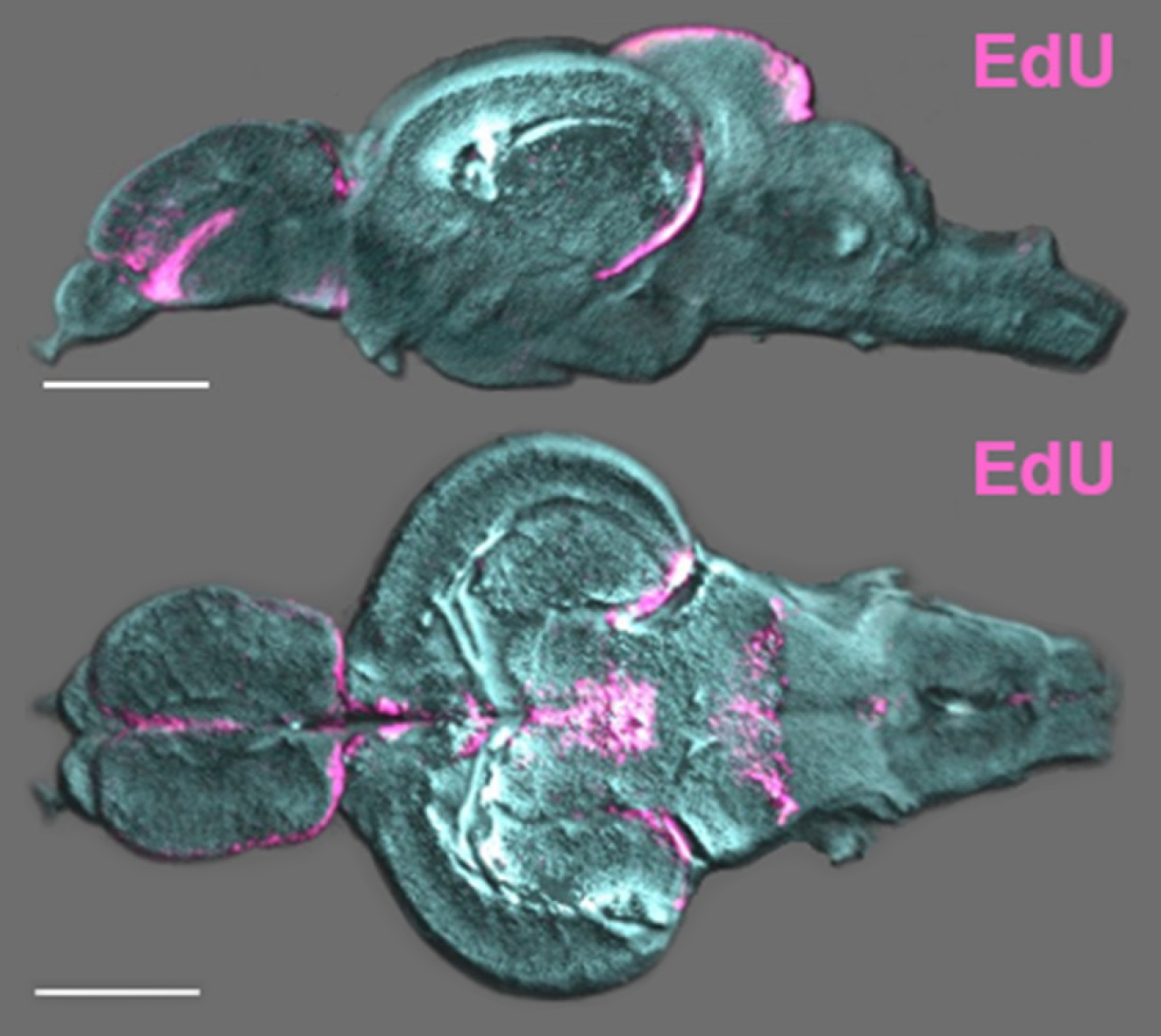
    [vc_row][vc_column][ultimate_spacer height=”35″ height_on_tabs=”25″ height_on_tabs_portrait=”25″ height_on_mob_landscape=”25″ height_on_mob=”25″][vc_custom_heading text=”FRONTIERS IN NEUROSCIENCE” font_container=”tag:p|text_align:left|line_height:1.3em” google_fonts=”font_family:Muli%3A300%2C300italic%2Cregular%2Citalic|font_style:400%20regular%3A400%3Anormal” css_animation=”slideInLeft”][ultimate_spacer height=”10″ height_on_tabs=”10″ height_on_tabs_portrait=”10″ height_on_mob_landscape=”10″ height_on_mob=”10″][vc_custom_heading text=”A Whole Brain Staining, Embedding, and Clearing Pipeline for Adult Zebrafish to Visualize Cell Proliferation and Morphology in 3-Dimensions” font_container=”tag:h3|text_align:left|line_height:1.3em” google_fonts=”font_family:Muli%3A300%2C300italic%2Cregular%2Citalic|font_style:400%20regular%3A400%3Anormal” css_animation=”slideInLeft”][ultimate_spacer height=”10″ height_on_tabs=”10″ height_on_tabs_portrait=”10″ height_on_mob_landscape=”10″ height_on_mob=”10″][vc_column_text]Benjamin W Lindsey, Alon M. Douek, Felix Loosli and Jan Kaslin[/vc_column_text][ultimate_spacer height=”25″ height_on_tabs=”25″ height_on_tabs_portrait=”25″ height_on_mob_landscape=”25″ height_on_mob=”25″][vc_column_text]The field of macro-imaging has grown considerably with the appearance of innovative clearing methods and confocal microscopes with lasers capable of penetrating increasing tissue depths. The ability to visualize and model the growth of whole organs as they develop from birth, or with manipulation, disease or injury, provides new ways of thinking about development, tissue-wide signaling, and cell-to-cell interactions. The zebrafish (Danio rerio) has ascended from a predominantly developmental model to a leading adult model of tissue regeneration. The unmatched neurogenic and regenerative capacity of the mature central nervous system, in particular, has received much attention, however tools to interrogate the adult brain are sparse. At present there exists no straightforward methods of visualizing changes in the whole adult brain in 3-dimensions (3-D) to examine systemic patterns of cell proliferation or cell populations of interest under physiological, injury, or diseased conditions. The method presented here is the first of its kind to offer an efficient step-by-step pipeline from intraperitoneal injections of the proliferative marker, 5-ethynyl-2′-deoxyuridine (EdU), to whole brain labeling, to a final embedded and cleared brain sample suitable for 3-D imaging using optical projection tomography (OPT). Moreover, this method allows potential for imaging GFP-reporter lines and cell-specific antibodies in the presence or absence of EdU. The small size of the adult zebrafish brain, the highly consistent degree of EdU labeling, and the use of basic clearing agents, benzyl benzoate, and benzyl alcohol, makes this method highly tractable for most laboratories interested in understanding the vertebrate central nervous system in health and disease. Post-processing of OPT-imaged adult zebrafish brains injected with EdU illustrate that proliferative patterns in EdU can readily be observed and analyzed using IMARIS and/or FIJI/IMAGEJ software. This protocol will be a valuable tool to unlock new ways of understanding systemic patterns in cell proliferation in the healthy and injured brain, brain-wide cellular interactions, stem cell niche development, and changes in brain morphology.[/vc_column_text][ultimate_spacer height=”25″ height_on_tabs=”25″ height_on_tabs_portrait=”25″ height_on_mob_landscape=”25″ height_on_mob=”25″][vc_column_text]READ FULL PUBLICATION

    Lindsey et al., 2018 (5.5 MB)

    [/vc_column_text][ultimate_spacer height=”25″ height_on_tabs=”25″ height_on_tabs_portrait=”25″ height_on_mob_landscape=”25″ height_on_mob=”25″][ultimate_spacer height=”25″ height_on_tabs=”25″ height_on_tabs_portrait=”25″ height_on_mob_landscape=”25″ height_on_mob=”25″][/vc_column][/vc_row][vc_row][vc_column][vc_btn title=”back to publications” color=”orange” i_icon_fontawesome=”fa fa-bookmark-o” add_icon=”true” link=”url:http%3A%2F%2Flindseylab.ca%2Fpublications%2F|title:PUBLICATIONS%202||”][/vc_column][/vc_row]

  • Optical Projection Tomography as a Novel Method to Visualize and Quantitate Whole-Brain Patterns of Cell Proliferation in the Adult Zebrafish Brain

    Optical Projection Tomography as a Novel Method to Visualize and Quantitate Whole-Brain Patterns of Cell Proliferation in the Adult Zebrafish Brain

    [vc_row][vc_column][ultimate_spacer height=”35″ height_on_tabs=”25″ height_on_tabs_portrait=”25″ height_on_mob_landscape=”25″ height_on_mob=”25″][vc_custom_heading text=”ZEBRAFISH, Volume 14, Number 6,” font_container=”tag:p|text_align:left|line_height:1.3em” google_fonts=”font_family:Muli%3A300%2C300italic%2Cregular%2Citalic|font_style:400%20regular%3A400%3Anormal” css_animation=”slideInLeft”][ultimate_spacer height=”10″ height_on_tabs=”10″ height_on_tabs_portrait=”10″ height_on_mob_landscape=”10″ height_on_mob=”10″][vc_custom_heading text=”Optical Projection Tomography as a Novel Method to Visualize and Quantitate Whole-Brain Patterns of Cell Proliferation in the Adult Zebrafish Brain” font_container=”tag:h3|text_align:left|line_height:1.3em” google_fonts=”font_family:Muli%3A300%2C300italic%2Cregular%2Citalic|font_style:400%20regular%3A400%3Anormal” css_animation=”slideInLeft”][ultimate_spacer height=”10″ height_on_tabs=”10″ height_on_tabs_portrait=”10″ height_on_mob_landscape=”10″ height_on_mob=”10″][vc_column_text]Benjamin W Lindsey and Jan Kaslin[/vc_column_text][ultimate_spacer height=”25″ height_on_tabs=”25″ height_on_tabs_portrait=”25″ height_on_mob_landscape=”25″ height_on_mob=”25″][vc_column_text]The zebrafish has become a popular model in neuroscience to study changes arising in the mature central nervous system (CNS) as a result of social behaviors, neurogenesis, brain injury and disorders, regeneration, neuroimmune interactions, and neurodegenerative diseases. Unfortunately, few brain-wide imaging tools are available to researchers investigating changes in the CNS during adulthood. Most studies continue to be performed by the standard method of fixing whole adult brains, sectioning, and confocal imaging antibody markers of interest. However, serial sectioning is time-consuming and severely limits interpretations of changes occurring across the entire brain axis. By contrast, the ability to visualize markers in threedimensional (3D) space allows investigators to screen the adult brain for regions of interest following manipulation. In this study, we have developed an efficient pipeline to clear, visualize, and quantitate the whole adult zebrafish brain using optical projection tomography (OPT) to assess changes in cell proliferation alone or in combination with transgenic reporter lines.[/vc_column_text][ultimate_spacer height=”25″ height_on_tabs=”25″ height_on_tabs_portrait=”25″ height_on_mob_landscape=”25″ height_on_mob=”25″][vc_column_text]READ FULL PUBLICATION

    Lindsey and Kaslin, 2017 (413 KB)

    [/vc_column_text][ultimate_spacer height=”25″ height_on_tabs=”25″ height_on_tabs_portrait=”25″ height_on_mob_landscape=”25″ height_on_mob=”25″][vc_btn title=”back to publications” color=”orange” i_icon_fontawesome=”fa fa-bookmark-o” add_icon=”true” link=”url:http%3A%2F%2Flindseylab.ca%2Fpublications%2F|title:PUBLICATIONS%202||”][ultimate_spacer height=”25″ height_on_tabs=”25″ height_on_tabs_portrait=”25″ height_on_mob_landscape=”25″ height_on_mob=”25″][/vc_column][/vc_row]
  • Mural lymphatic endothelial cells regulate meningeal angiogenesis in the zebrafish

    Mural lymphatic endothelial cells regulate meningeal angiogenesis in the zebrafish

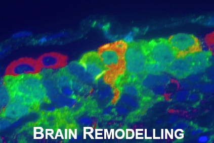
    [vc_row][vc_column][ultimate_spacer height=”35″ height_on_tabs=”25″ height_on_tabs_portrait=”25″ height_on_mob_landscape=”25″ height_on_mob=”25″][vc_custom_heading text=”NATURE NEUROSCIENCE” font_container=”tag:p|text_align:left|line_height:1.3em” google_fonts=”font_family:Muli%3A300%2C300italic%2Cregular%2Citalic|font_style:400%20regular%3A400%3Anormal” css_animation=”slideInLeft”][ultimate_spacer height=”10″ height_on_tabs=”10″ height_on_tabs_portrait=”10″ height_on_mob_landscape=”10″ height_on_mob=”10″][vc_custom_heading text=”Mural lymphatic endothelial cells regulate meningeal angiogenesis in the zebrafish” font_container=”tag:h3|text_align:left|line_height:1.3em” google_fonts=”font_family:Muli%3A300%2C300italic%2Cregular%2Citalic|font_style:400%20regular%3A400%3Anormal” css_animation=”slideInLeft”][ultimate_spacer height=”10″ height_on_tabs=”10″ height_on_tabs_portrait=”10″ height_on_mob_landscape=”10″ height_on_mob=”10″][vc_column_text]Neil I Bower, Katarzyna Koltowska, Cathy Pichol-Thievend, Isaac Virshup, Scott Paterson, Anne K Lagendijk, Weili Wang, Benjamin W Lindsey, Stephen J Bent, Sungmin Baek, Maria Rondon-Galeano, Daniel G Hurley, Naoki Mochizuki, Cas Simons, Mathias Francois, Christine A Wells , Jan Kaslin & Benjamin M Hogan [/vc_column_text][ultimate_spacer height=”25″ height_on_tabs=”25″ height_on_tabs_portrait=”25″ height_on_mob_landscape=”25″ height_on_mob=”25″][vc_column_text]Mural cells of the vertebrate brain maintain vascular integrity and function, play roles in stroke and are involved in maintenance of neural stem cells. However, the origins, diversity and roles of mural cells remain to be fully understood. Using transgenic zebrafish, we identified a population of isolated mural lymphatic endothelial cells surrounding meningeal blood vessels. These meningeal mural lymphatic endothelial cells (muLECs) express lymphatic endothelial cell markers and form by sprouting from blood vessels. In larvae, muLECs develop from a lymphatic endothelial loop in the midbrain into a dispersed, nonlumenized mural lineage. muLEC development requires normal signaling through the Vegfc–Vegfd–Ccbe1–Vegfr3 pathway. Mature muLECs produce vascular growth factors and accumulate low-density lipoproteins from the bloodstream. We find that muLECs are essential for normal meningeal vascularization. Together, these data identify an unexpected lymphatic lineage and developmental mechanism necessary for establishing normal meningeal blood vasculature.[/vc_column_text][ultimate_spacer height=”25″ height_on_tabs=”25″ height_on_tabs_portrait=”25″ height_on_mob_landscape=”25″ height_on_mob=”25″][vc_column_text]READ FULL PUBLICATION

    Bower et al., 2017 (1.3 MB)

    Bower et al., 2017 supplementary (2 MB)[/vc_column_text][ultimate_spacer height=”25″ height_on_tabs=”25″ height_on_tabs_portrait=”25″ height_on_mob_landscape=”25″ height_on_mob=”25″][vc_btn title=”back to publications” color=”orange” i_icon_fontawesome=”fa fa-bookmark-o” add_icon=”true” link=”url:http%3A%2F%2Flindseylab.ca%2Fpublications%2F|title:PUBLICATIONS%202||”][ultimate_spacer height=”25″ height_on_tabs=”25″ height_on_tabs_portrait=”25″ height_on_mob_landscape=”25″ height_on_mob=”25″][/vc_column][/vc_row]
  • A library of AuNPs modified by RAFT polymers of different charge and chain length

    A library of AuNPs modified by RAFT polymers of different charge and chain length

    [vc_row][vc_column][ultimate_spacer height=”35″ height_on_tabs=”25″ height_on_tabs_portrait=”25″ height_on_mob_landscape=”25″ height_on_mob=”25″][vc_custom_heading text=”ROYAL SOCIETY OF CHEMISTRY” font_container=”tag:p|text_align:left|line_height:1.3em” google_fonts=”font_family:Muli%3A300%2C300italic%2Cregular%2Citalic|font_style:400%20regular%3A400%3Anormal” css_animation=”slideInLeft”][ultimate_spacer height=”10″ height_on_tabs=”10″ height_on_tabs_portrait=”10″ height_on_mob_landscape=”10″ height_on_mob=”10″][vc_custom_heading text=”A library of AuNPs modified by RAFT polymers of different charge and chain length: high throughput synthesis and synchrotron XFM imaging using a zebrafish larvae model” font_container=”tag:h3|text_align:left|line_height:1.3em” google_fonts=”font_family:Muli%3A300%2C300italic%2Cregular%2Citalic|font_style:400%20regular%3A400%3Anormal” css_animation=”slideInLeft”][ultimate_spacer height=”10″ height_on_tabs=”10″ height_on_tabs_portrait=”10″ height_on_mob_landscape=”10″ height_on_mob=”10″][vc_column_text]Bao Luan, Timo Friedrich, Jiali Zhai, Victor A. Streltsov, Benjamin W Lindsey, Jan Kaslin, Martin D. de Jonge, Jin Zhu, Timothy C. Hughes and Xiaojuan Hao[/vc_column_text][ultimate_spacer height=”25″ height_on_tabs=”25″ height_on_tabs_portrait=”25″ height_on_mob_landscape=”25″ height_on_mob=”25″][vc_column_text]Gold nanoparticles (AuNPs) have been widely investigated in drug delivery and imaging. However, for such biomedical applications, the modification of AuNPs is necessary to improve their aqueous dispersion stability and biocompatibility, especially in a salt environment. Here, we report a simple and highly efficient method to create a library of polymer-modified gold nanoparticles (PAuNPs) and screen their dispersion stability utilizing high-throughput facilities (in total 1000 experiments). Three types of water soluble polymers with different charge and chain length were prepared using Reversible Addition-Fragmentation chain Transfer (RAFT) polymerization technology. The polymers were then converted into thiol-capped polymers by aminolysis and coated on AuNPs via thiol–gold binding to improve aqueous dispersion stability as well as the biocompatibility of AuNPs. Remarkably, we present the first report of imaging zebrafish embryos injected with a PAuNPs sample (selected from our PAuNPs library) using synchrotron X-ray fluorescence microscopy (XFM) beamline. The selected PAuNPs sample, which has been evaluated to be non-cytotoxic to L929 cells and biocompatible to zebrafish larvae in a wide range of concentrations, was injected into zebrafish larvae via the cardinal vein and could be clearly visualized in the whole circulatory system including both peripheral blood vessels and the head region by XFM. The result indicates that zebrafish larvae could be a potential animal model for probing the ability of AuNPs to cross the blood–brain barrier (BBB) and therefore hold promise for investigating AuNPs in biomedical applications such as detecting cancer and Alzheimer’s disease (AD).[/vc_column_text][ultimate_spacer height=”25″ height_on_tabs=”25″ height_on_tabs_portrait=”25″ height_on_mob_landscape=”25″ height_on_mob=”25″][vc_column_text]READ FULL PUBLICATION

    Luan et al., 2016 (950 KB)

    [/vc_column_text][ultimate_spacer height=”25″ height_on_tabs=”25″ height_on_tabs_portrait=”25″ height_on_mob_landscape=”25″ height_on_mob=”25″][vc_btn title=”back to publications” color=”orange” i_icon_fontawesome=”fa fa-bookmark-o” add_icon=”true” link=”url:http%3A%2F%2Flindseylab.ca%2Fpublications%2F|title:PUBLICATIONS%202||”][ultimate_spacer height=”25″ height_on_tabs=”25″ height_on_tabs_portrait=”25″ height_on_mob_landscape=”25″ height_on_mob=”25″][/vc_column][/vc_row]
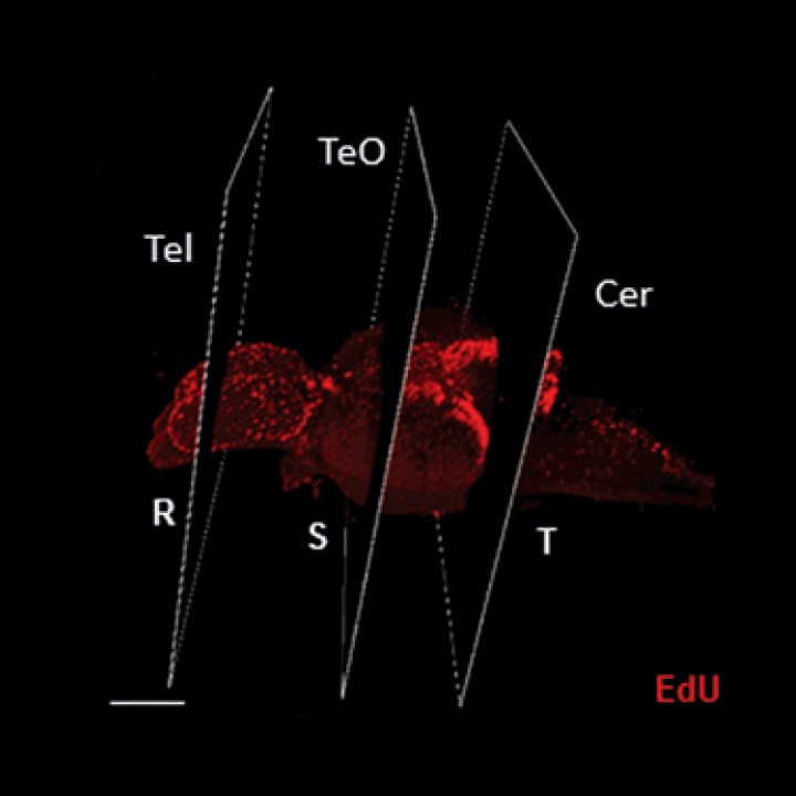
  • Changes in the Social Environment Induce Neurogenic Plasticity Predominantly in Niches Residing in Sensory Structures of the Zebrafish Brain Independently of Cortisol Levels

    Changes in the Social Environment Induce Neurogenic Plasticity Predominantly in Niches Residing in Sensory Structures of the Zebrafish Brain Independently of Cortisol Levels

    [vc_row][vc_column][ultimate_spacer height=”35″ height_on_tabs=”25″ height_on_tabs_portrait=”25″ height_on_mob_landscape=”25″ height_on_mob=”25″][vc_custom_heading text=”WILEY PERIODICALS, INC.” font_container=”tag:p|text_align:left|line_height:1.3em” google_fonts=”font_family:Muli%3A300%2C300italic%2Cregular%2Citalic|font_style:400%20regular%3A400%3Anormal” css_animation=”slideInLeft”][ultimate_spacer height=”10″ height_on_tabs=”10″ height_on_tabs_portrait=”10″ height_on_mob_landscape=”10″ height_on_mob=”10″][vc_custom_heading text=”Changes in the Social Environment Induce Neurogenic Plasticity Predominantly in Niches Residing in Sensory Structures of the Zebrafish Brain Independently of Cortisol Levels” font_container=”tag:h3|text_align:left|line_height:1.3em” google_fonts=”font_family:Muli%3A300%2C300italic%2Cregular%2Citalic|font_style:400%20regular%3A400%3Anormal” css_animation=”slideInLeft”][ultimate_spacer height=”10″ height_on_tabs=”10″ height_on_tabs_portrait=”10″ height_on_mob_landscape=”10″ height_on_mob=”10″][vc_column_text]Benjamin W Lindsey and Vincent Tropepe [/vc_column_text][ultimate_spacer height=”25″ height_on_tabs=”25″ height_on_tabs_portrait=”25″ height_on_mob_landscape=”25″ height_on_mob=”25″][vc_column_text]The social environment is known to regulate constitutive rates of adult neurogenesis in vertebrates, potentially influencing stem/progenitor proliferation, survival of the post-mitotic population, or the extent of neuronal differentiation (Lindsey and Tropepe, 2006; Kempermann, 2011; Maruska et al., 2012). By deconstructing the complexity of the social environment, however, it is clear that much of the information that an animal encodes encompasses both learned components that are processed in higherorder brain centers of the forebrain, such as the hippocampus and neocortex, as well as modality-specific sensory components that are processed foremost in corresponding primary sensory structures. What happens then to constitutive rates of adult neurogenesis when social animals venture outside of their home range, are separated from their social group, or undergo novel social interactions? How might environmental change activate neurogenic plasticity and alter the composition of the neurogenic niche in the adult vertebrate brain? To begin to address these questions, neurogenic niches residing within primary sensory structures of the brain must be compared with those localized to higher-order forebrain structures to determine if there are differential responses at the cellular level to deviations from a familiar social context.[/vc_column_text][ultimate_spacer height=”25″ height_on_tabs=”25″ height_on_tabs_portrait=”25″ height_on_mob_landscape=”25″ height_on_mob=”25″][vc_column_text]READ FULL PUBLICATION

    Lindsey and Tropepe, 2006 (1.5 MB)

    [/vc_column_text][ultimate_spacer height=”25″ height_on_tabs=”25″ height_on_tabs_portrait=”25″ height_on_mob_landscape=”25″ height_on_mob=”25″][vc_btn title=”back to publications” color=”orange” i_icon_fontawesome=”fa fa-bookmark-o” add_icon=”true” link=”url:http%3A%2F%2Flindseylab.ca%2Fpublications%2F|title:PUBLICATIONS%202||”][ultimate_spacer height=”25″ height_on_tabs=”25″ height_on_tabs_portrait=”25″ height_on_mob_landscape=”25″ height_on_mob=”25″][/vc_column][/vc_row]
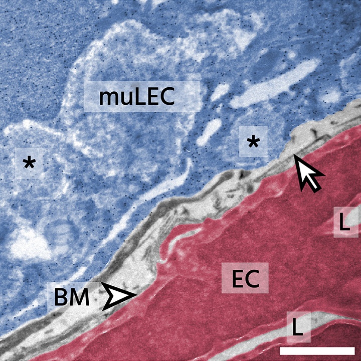
  • Sensory-specific modulation of adult neurogenesis in sensory structures is associated with the type of stem cell present in the neurogenic niche of the zebrafish brain

    Sensory-specific modulation of adult neurogenesis in sensory structures is associated with the type of stem cell present in the neurogenic niche of the zebrafish brain

    [vc_row][vc_column][ultimate_spacer height=”35″ height_on_tabs=”25″ height_on_tabs_portrait=”25″ height_on_mob_landscape=”25″ height_on_mob=”25″][vc_custom_heading text=”EUROPEAN JOURNAL OF NEUROSCIENCE” font_container=”tag:p|text_align:left|line_height:1.3em” google_fonts=”font_family:Muli%3A300%2C300italic%2Cregular%2Citalic|font_style:400%20regular%3A400%3Anormal” css_animation=”slideInLeft”][ultimate_spacer height=”10″ height_on_tabs=”10″ height_on_tabs_portrait=”10″ height_on_mob_landscape=”10″ height_on_mob=”10″][vc_custom_heading text=”Sensory-specific modulation of adult neurogenesis in sensory structures is associated with the type of stem cell present in the neurogenic niche of the zebrafish brain” font_container=”tag:h3|text_align:left|line_height:1.3em” google_fonts=”font_family:Muli%3A300%2C300italic%2Cregular%2Citalic|font_style:400%20regular%3A400%3Anormal” css_animation=”slideInLeft”][ultimate_spacer height=”10″ height_on_tabs=”10″ height_on_tabs_portrait=”10″ height_on_mob_landscape=”10″ height_on_mob=”10″][vc_column_text]Benjamin W Lindsey, Sabrina Di Donato, Jan Kaslin and Vincent Tropepe[/vc_column_text][ultimate_spacer height=”25″ height_on_tabs=”25″ height_on_tabs_portrait=”25″ height_on_mob_landscape=”25″ height_on_mob=”25″][vc_column_text]Vertebrate adult neurogenesis can be modulated through changes in proliferation, survival or differentiation, and this appears to be associated with distinct functional requirements for new neurons (Grandel & Brand, 2013). Outside of mammals and birds however, there is little understanding of how adult neurogenesis may function as a biological substrate to modify species behaviour or information processing.

    Studies of neurogenesis in the adult zebrafish forebrain have demonstrated that the cell types within neurogenic niches consist of populations of proliferative and non-proliferative glia that can be classified using immunohistochemical markers, with the stem/progenitor phenotype commonly having a radial glial (RG) profile (Ganz et al., 2010; Marz et al., 2010). Transmission electron microscopy (TEM) studies have revealed that the composition of different adult niches is further determined by the presence and frequency of seven distinct cell types (Type IIa–Type VI), with pallial niches characterised by Type IIa RG-like cells at the ventricular surface and subpallial niches composed of layers of elongated Type III cells reminiscent of neuroepithelial (NE)-like profiles (Lindsey et al., 2012). Accordingly, it has been documented that the cerebellum and optic tectum (TeO) also retain stem/progenitors with NE features similar to early development (Kaslin et al., 2009, 2013; Alunni et al., 2010; Ito et al., 2010), though ultrastructural evidence for this is lacking.[/vc_column_text][ultimate_spacer height=”25″ height_on_tabs=”25″ height_on_tabs_portrait=”25″ height_on_mob_landscape=”25″ height_on_mob=”25″][vc_column_text]READ FULL PUBLICATION

    Lindsey et al., 2014 (954 KB)

    [/vc_column_text][ultimate_spacer height=”25″ height_on_tabs=”25″ height_on_tabs_portrait=”25″ height_on_mob_landscape=”25″ height_on_mob=”25″][vc_btn title=”back to publications” color=”orange” i_icon_fontawesome=”fa fa-bookmark-o” add_icon=”true” link=”url:http%3A%2F%2Flindseylab.ca%2Fpublications%2F|title:PUBLICATIONS%202||”][ultimate_spacer height=”25″ height_on_tabs=”25″ height_on_tabs_portrait=”25″ height_on_mob_landscape=”25″ height_on_mob=”25″][/vc_column][/vc_row]
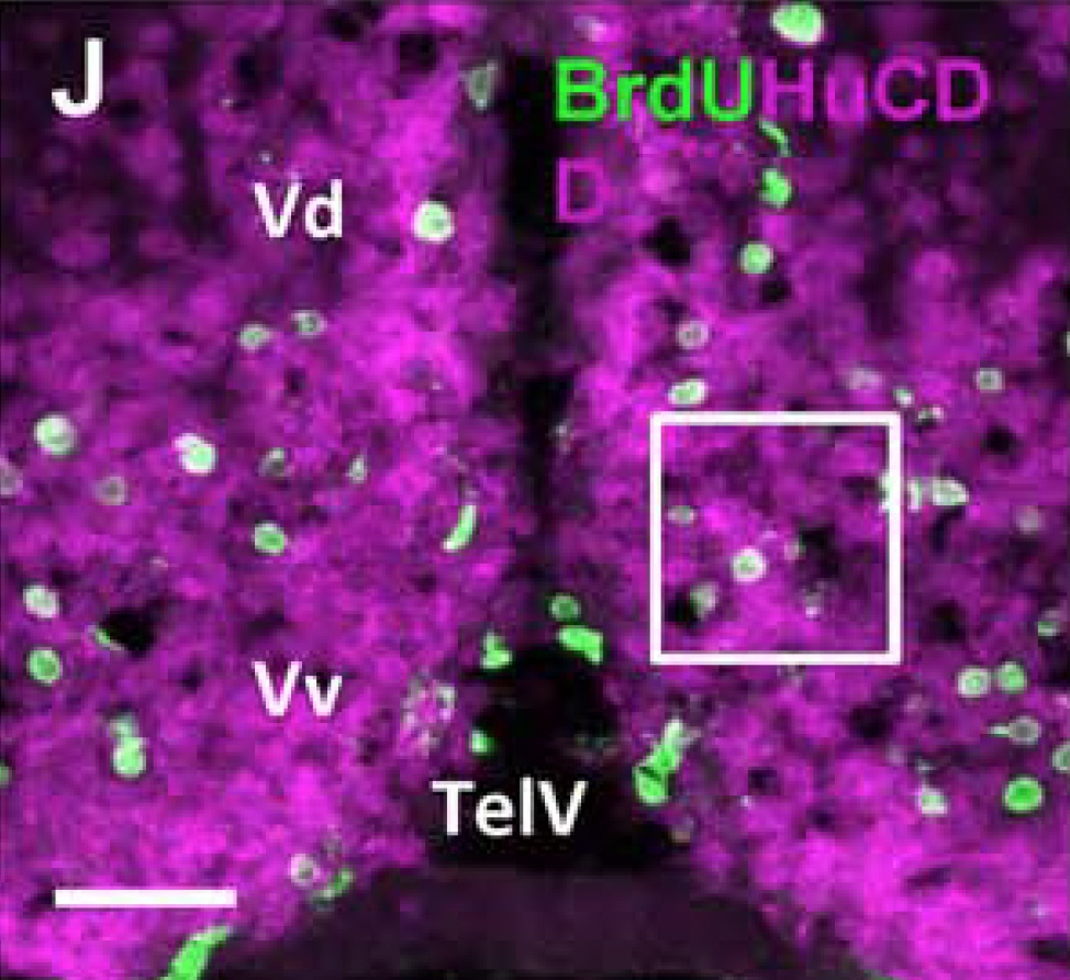
  • The Cellular Composition of Neurogenic Periventricular Zones in the Adult Zebrafish Forebrain

    The Cellular Composition of Neurogenic Periventricular Zones in the Adult Zebrafish Forebrain

    [vc_row][vc_column][ultimate_spacer height=”35″ height_on_tabs=”25″ height_on_tabs_portrait=”25″ height_on_mob_landscape=”25″ height_on_mob=”25″][vc_custom_heading text=”WILEY PERIODICALS, INC.” font_container=”tag:p|text_align:left|line_height:1.3em” google_fonts=”font_family:Muli%3A300%2C300italic%2Cregular%2Citalic|font_style:400%20regular%3A400%3Anormal” css_animation=”slideInLeft”][ultimate_spacer height=”10″ height_on_tabs=”10″ height_on_tabs_portrait=”10″ height_on_mob_landscape=”10″ height_on_mob=”10″][vc_custom_heading text=”The Cellular Composition of Neurogenic Periventricular Zones in the Adult Zebrafish Forebrain” font_container=”tag:h3|text_align:left|line_height:1.3em” google_fonts=”font_family:Muli%3A300%2C300italic%2Cregular%2Citalic|font_style:400%20regular%3A400%3Anormal” css_animation=”slideInLeft”][ultimate_spacer height=”10″ height_on_tabs=”10″ height_on_tabs_portrait=”10″ height_on_mob_landscape=”10″ height_on_mob=”10″][vc_column_text]Benjamin W Lindsey, Audrey Darabie, and Vincent Tropepe[/vc_column_text][ultimate_spacer height=”25″ height_on_tabs=”25″ height_on_tabs_portrait=”25″ height_on_mob_landscape=”25″ height_on_mob=”25″][vc_column_text]Adult neurogenesis occurs in well-circumscribed regions of the mature vertebrate brain. This process is initiated by the proliferation of stem and/or progenitor cells, which over time generate new neurons and glia. Adult neurogenesis is regulated, in part, through the interaction between neurogenic cells and neighboring cells that are present within their immediate microenvironment, or niche. Moreover, the niche itself may be largely unique in terms of its cellular composition, degree of plasticity, and the molecular factors maintaining it as an active site of adult neurogenesis in different regions of the brain or at different times during development. For instance, studies in the adult rodent brain show differences in the time course of neuronal differentiation, and the neuronal subtypes that arise from the subependymal zone (SEZ; also referred to as the subventricular zone [SVZ]) of the forebrain lateral ventricles or the subgranular zone (SGZ) of the hippocampal dentate gyrus (reviewed in Zhao et al., 2008).

    These findings support the notion of niche-specific regulation at the cellular and molecular levels. Despite significant advances using rodent models, many nonmammalian vertebrates offer the opportunity to explore the regulation and composition of the neurogenic niche in both homologous structures as well as within novel neuroanatomical loci where this trait is absent in mammals (Zupanc, 2001). Understanding how adult neurogenic niches are differentially regulated across vertebrate species will be an essential step in gaining a fundamental understanding of the functional significance of this conserved trait (Lindsey and Tropepe, 2006).[/vc_column_text][ultimate_spacer height=”25″ height_on_tabs=”25″ height_on_tabs_portrait=”25″ height_on_mob_landscape=”25″ height_on_mob=”25″][vc_column_text]READ FULL PUBLICATION

    Lindsey et al., 2012 (2 MB)

    [/vc_column_text][ultimate_spacer height=”25″ height_on_tabs=”25″ height_on_tabs_portrait=”25″ height_on_mob_landscape=”25″ height_on_mob=”25″][vc_btn title=”back to publications” color=”orange” i_icon_fontawesome=”fa fa-bookmark-o” add_icon=”true” link=”url:http%3A%2F%2Flindseylab.ca%2Fpublications%2F|title:PUBLICATIONS%202||”][ultimate_spacer height=”25″ height_on_tabs=”25″ height_on_tabs_portrait=”25″ height_on_mob_landscape=”25″ height_on_mob=”25″][/vc_column][/vc_row]
  • Effects of Simulated Microgravity on the Development of the Swimbladder and Buoyancy Control in Larval Zebrafish (Danio rerio)

    Effects of Simulated Microgravity on the Development of the Swimbladder and Buoyancy Control in Larval Zebrafish (Danio rerio)

    [vc_row][vc_column][ultimate_spacer height=”35″ height_on_tabs=”25″ height_on_tabs_portrait=”25″ height_on_mob_landscape=”25″ height_on_mob=”25″][vc_custom_heading text=”JOURNAL OF EXPERIMENTAL ZOOLOGY” font_container=”tag:p|text_align:left|line_height:1.3em” google_fonts=”font_family:Muli%3A300%2C300italic%2Cregular%2Citalic|font_style:400%20regular%3A400%3Anormal” css_animation=”slideInLeft”][ultimate_spacer height=”10″ height_on_tabs=”10″ height_on_tabs_portrait=”10″ height_on_mob_landscape=”10″ height_on_mob=”10″][vc_custom_heading text=”Effects of Simulated Microgravity on the Development of the Swimbladder and Buoyancy Control in Larval Zebrafish (Danio rerio)” font_container=”tag:h3|text_align:left|line_height:1.3em” google_fonts=”font_family:Muli%3A300%2C300italic%2Cregular%2Citalic|font_style:400%20regular%3A400%3Anormal” css_animation=”slideInLeft”][ultimate_spacer height=”10″ height_on_tabs=”10″ height_on_tabs_portrait=”10″ height_on_mob_landscape=”10″ height_on_mob=”10″][vc_column_text]Benjamin W Lindsey, Tristan C. Dumbarton, Stephen J. Moorman, Frank M. Smith, and Roger P. Croll[/vc_column_text][ultimate_spacer height=”25″ height_on_tabs=”25″ height_on_tabs_portrait=”25″ height_on_mob_landscape=”25″ height_on_mob=”25″][vc_column_text]The gas-filled swimbladder of teleost fishes provides hydrodynamic lift which counteracts the high density of other body tissues, and thereby allows the fish to achieve neutral buoyancy with minimal energy expenditure. In this study, we examined whether the absence of a constant direction gravitational vector affects the ontogeny of the swimbladder and buoyancy control in zebrafish (Danio rerio). We exposed fertilized eggs to simulated microgravity (SMG) in a closed rotating wall vessel with control eggs placed in a similar but nonrotating container. All eggs hatched in both groups. At 96 hr of postfertilization (hpf), all larvae were removed from the experimental and control vessels. At this point, 62% of the control larvae, but only 14% of SMG-exposed larvae, were observed to have inflated their swimbladder. In addition, the mean volume of the inflated swimbladders was significantly greater in the control larvae compared with larvae raised in SMG. After transfer to open stationary observation tanks, larvae with uninflated swimbladders in both groups swam to the surface to complete inflation, but this process was significantly delayed in larvae exposed to SMG. Initial differences in swimbladder inflation and volume between groups disappeared by 144 hpf. Furthermore, there were no apparent changes in patterns of development and maturation of swimbladder musculature, vasculature, or innervation resulting from SMG exposure at later stages of ontogeny. These data indicate that, despite a transient delay in swimbladder inflation in zebrafish larvae exposed to SMG, subsequent swimbladder development in these animals proceeded similarly to that in normal larvae.[/vc_column_text][ultimate_spacer height=”25″ height_on_tabs=”25″ height_on_tabs_portrait=”25″ height_on_mob_landscape=”25″ height_on_mob=”25″][vc_column_text]READ FULL PUBLICATION

    Lindsey et al., 2011 (458 KB)

    [/vc_column_text][ultimate_spacer height=”25″ height_on_tabs=”25″ height_on_tabs_portrait=”25″ height_on_mob_landscape=”25″ height_on_mob=”25″][vc_btn title=”back to publications” color=”orange” i_icon_fontawesome=”fa fa-bookmark-o” add_icon=”true” link=”url:http%3A%2F%2Flindseylab.ca%2Fpublications%2F|title:PUBLICATIONS%202||”][ultimate_spacer height=”25″ height_on_tabs=”25″ height_on_tabs_portrait=”25″ height_on_mob_landscape=”25″ height_on_mob=”25″][/vc_column][/vc_row]
  • From Inflation to Flotation: Contribution of the Swimbladder to Whole-Body Density and Swimming Depth During Development of the Zebrafish (Danio rerio)

    From Inflation to Flotation: Contribution of the Swimbladder to Whole-Body Density and Swimming Depth During Development of the Zebrafish (Danio rerio)

    [vc_row][vc_column][ultimate_spacer height=”35″ height_on_tabs=”25″ height_on_tabs_portrait=”25″ height_on_mob_landscape=”25″ height_on_mob=”25″][vc_custom_heading text=”ZEBRAFISH, Volume 7, Number 1″ font_container=”tag:p|text_align:left|line_height:1.3em” google_fonts=”font_family:Muli%3A300%2C300italic%2Cregular%2Citalic|font_style:400%20regular%3A400%3Anormal” css_animation=”slideInLeft”][ultimate_spacer height=”10″ height_on_tabs=”10″ height_on_tabs_portrait=”10″ height_on_mob_landscape=”10″ height_on_mob=”10″][vc_custom_heading text=”From Inflation to Flotation: Contribution of the Swimbladder to Whole-Body Density and Swimming Depth During Development of the Zebrafish (Danio rerio)” font_container=”tag:h3|text_align:left|line_height:1.3em” google_fonts=”font_family:Muli%3A300%2C300italic%2Cregular%2Citalic|font_style:400%20regular%3A400%3Anormal” css_animation=”slideInLeft”][ultimate_spacer height=”10″ height_on_tabs=”10″ height_on_tabs_portrait=”10″ height_on_mob_landscape=”10″ height_on_mob=”10″][vc_column_text]Benjamin W Lindsey, Frank M. Smith, and Roger P. Croll[/vc_column_text][ultimate_spacer height=”25″ height_on_tabs=”25″ height_on_tabs_portrait=”25″ height_on_mob_landscape=”25″ height_on_mob=”25″][vc_column_text]Teleost fish contain a variety of tissues, such as muscle, cartilage, and bone, which are denser than the external aqueous environment. Without compensatory mechanisms to provide buoyancy, fish would, therefore, sink. Consequently, the performance of essential behaviors such as feeding, predator avoidance, migrations, and reproduction1 would be energetically costly as fish would need to swim constantly to maintain a vertical position in the water column. To overcome this problem, fish have evolved numerous mechanisms to achieve neutral buoyancy. These mechanisms include the development of watery muscle, synthesis of low-density lipids, and reduction in bone mass (reviewed in Refs.1–3). However, of all buoyancy compensating mechanisms, a gasfilled organ in the coelomic cavity, the swimbladder, is considered to be the most efficient.2 By controlling the volume of gas in the swimbladder, fish can attain neutral buoyancy at any depth, thereby minimizing the amount of energy expended by swimming to hold vertical station in the water column.[/vc_column_text][ultimate_spacer height=”25″ height_on_tabs=”25″ height_on_tabs_portrait=”25″ height_on_mob_landscape=”25″ height_on_mob=”25″][vc_column_text]READ FULL PUBLICATION

    Lindsey et al., 2010 (3.1 MB)

    [/vc_column_text][ultimate_spacer height=”25″ height_on_tabs=”25″ height_on_tabs_portrait=”25″ height_on_mob_landscape=”25″ height_on_mob=”25″][vc_btn title=”back to publications” color=”orange” i_icon_fontawesome=”fa fa-bookmark-o” add_icon=”true” link=”url:http%3A%2F%2Flindseylab.ca%2Fpublications%2F|title:PUBLICATIONS%202||”][ultimate_spacer height=”25″ height_on_tabs=”25″ height_on_tabs_portrait=”25″ height_on_mob_landscape=”25″ height_on_mob=”25″][/vc_column][/vc_row]
  • The Contribution of the Swimbladder to Buoyancy in the Adult Zebrafish (Danio rerio): A Morphometric Analysis

    The Contribution of the Swimbladder to Buoyancy in the Adult Zebrafish (Danio rerio): A Morphometric Analysis

    [vc_row][vc_column][ultimate_spacer height=”35″ height_on_tabs=”25″ height_on_tabs_portrait=”25″ height_on_mob_landscape=”25″ height_on_mob=”25″][vc_custom_heading text=”JOURNAL OF MORPHOLOGY” font_container=”tag:p|text_align:left|line_height:1.3em” google_fonts=”font_family:Muli%3A300%2C300italic%2Cregular%2Citalic|font_style:400%20regular%3A400%3Anormal” css_animation=”slideInLeft”][ultimate_spacer height=”10″ height_on_tabs=”10″ height_on_tabs_portrait=”10″ height_on_mob_landscape=”10″ height_on_mob=”10″][vc_custom_heading text=”The Contribution of the Swimbladder to Buoyancy in the Adult Zebrafish (Danio rerio): A Morphometric Analysis” font_container=”tag:h3|text_align:left|line_height:1.3em” google_fonts=”font_family:Muli%3A300%2C300italic%2Cregular%2Citalic|font_style:400%20regular%3A400%3Anormal” css_animation=”slideInLeft”][ultimate_spacer height=”10″ height_on_tabs=”10″ height_on_tabs_portrait=”10″ height_on_mob_landscape=”10″ height_on_mob=”10″][vc_column_text]George N. Robertson, Benjamin W Lindsey, Tristan C. Dumbarton, Roger P. Croll, and Frank M. Smith[/vc_column_text][ultimate_spacer height=”25″ height_on_tabs=”25″ height_on_tabs_portrait=”25″ height_on_mob_landscape=”25″ height_on_mob=”25″][vc_column_text]A significant portion of the body mass of teleost fishes is composed of tissues such as bone and muscle that are denser than water (Alexander, 1993). Consequently, compensatory mechanisms are required to reduce the overall density of the body, in order to decrease the energetic cost of swimming to maintain vertical position in the water column. Pelagic teleosts have evolved several strategies to overcome the inherent negative buoyancy related to the density of body tissues. These include the synthesis of large amounts of lipid, the development of ‘‘watery’’ muscles, the reduction of bone mass, and the presence of an internal gas-filled chamber, the swimbladder (Alexander, 1972, 1989; Lefrancois et al., 2001). Of all the mechanisms used by fish to reduce total body density, the swimbladder has been proposed to be the most energy efficient (Alexander, 1993). The volume of low-density gas in the swimbladder offsets the higher density of body tissues so that fish possessing swimbladders are very close to neutral buoyancy at a specific depth in the water column. However, the quantitative contribution of the swimbladder to the attainment of neutral buoyancy has been established for only a few species of teleosts, including some cyprinids (Alexander, 1959) and the toadfish (Fine et al., 1995). Here we investigated the contribution of the swimbladder to buoyancy in a small fresh-water cyprinid, the zebrafish (Danio rerio).[/vc_column_text][ultimate_spacer height=”25″ height_on_tabs=”25″ height_on_tabs_portrait=”25″ height_on_mob_landscape=”25″ height_on_mob=”25″][vc_column_text]READ FULL PUBLICATION

    Robertson et al., 2008 (421 KB)

    [/vc_column_text][ultimate_spacer height=”25″ height_on_tabs=”25″ height_on_tabs_portrait=”25″ height_on_mob_landscape=”25″ height_on_mob=”25″][vc_btn title=”back to publications” color=”orange” i_icon_fontawesome=”fa fa-bookmark-o” add_icon=”true” link=”url:http%3A%2F%2Flindseylab.ca%2Fpublications%2F|title:PUBLICATIONS%202||”][ultimate_spacer height=”25″ height_on_tabs=”25″ height_on_tabs_portrait=”25″ height_on_mob_landscape=”25″ height_on_mob=”25″][/vc_column][/vc_row]
  • Easy and Rapid Differentiation of Embryonic Stem Cells into Functional Motoneurons Using Sonic Hedgehog-Producing Cells

    Easy and Rapid Differentiation of Embryonic Stem Cells into Functional Motoneurons Using Sonic Hedgehog-Producing Cells

    [vc_row][vc_column][ultimate_spacer height=”35″ height_on_tabs=”25″ height_on_tabs_portrait=”25″ height_on_mob_landscape=”25″ height_on_mob=”25″][vc_custom_heading text=”STEM CELLS” font_container=”tag:p|text_align:left|line_height:1.3em” google_fonts=”font_family:Muli%3A300%2C300italic%2Cregular%2Citalic|font_style:400%20regular%3A400%3Anormal” css_animation=”slideInLeft”][ultimate_spacer height=”10″ height_on_tabs=”10″ height_on_tabs_portrait=”10″ height_on_mob_landscape=”10″ height_on_mob=”10″][vc_custom_heading text=”Easy and Rapid Differentiation of Embryonic Stem Cells into Functional Motoneurons Using Sonic Hedgehog-Producing Cells” font_container=”tag:h3|text_align:left|line_height:1.3em” google_fonts=”font_family:Muli%3A300%2C300italic%2Cregular%2Citalic|font_style:400%20regular%3A400%3Anormal” css_animation=”slideInLeft”][ultimate_spacer height=”10″ height_on_tabs=”10″ height_on_tabs_portrait=”10″ height_on_mob_landscape=”10″ height_on_mob=”10″][vc_column_text]Prabakaran Soundararajan, Benjamin W Lindsey, Cindee Leopold and Victor F. Rafuse[/vc_column_text][ultimate_spacer height=”25″ height_on_tabs=”25″ height_on_tabs_portrait=”25″ height_on_mob_landscape=”25″ height_on_mob=”25″][vc_column_text]Spinal cord disorders such as amyotrophic lateral sclerosis (ALS) and spinal muscular atrophy result in the dysfunction and eventual death of spinal motoneurons. Both neurodegenerative disorders have been targeted for cell replacement therapy using stem cells because they involve individual classes of neurons that are located within defined regions of the central nervous system [1–3]. To this end, several investigators have successfully directed embryonic stem (ES) cells to differentiate into motoneurons in vitro [4–6]. Furthermore, recent studies have shown that transplantation of ES cell-derived motoneurons dramatically attenuates abnormal locomotor behavior in an animal model of ALS [7]. However, despite these promising results, substantially more research is required before ES cells can be used for clinical therapies. Consequently, protocols directing ES cells to differentiate into motoneurons should use methods that are readily available to a wide range of investigators with varying scientific backgrounds and technical expertise.

    The majority of protocols directing ES cells to differentiate into specific neuronal subtypes use a sequential combination of mitogens [8]. Unfortunately, some differentiation protocols use techniques or specific reagents that limit their widespread use. For example, a few laboratories have shown that ES cells readily differentiate into functional motoneurons when cultured with a sonic hedgehog (Shh) agonist (HhAg1.3) and retinoic acid (RA) [4, 5, 7, 9, 10]. Unfortunately, expansion of these studies by other investigators has been limited because the Shh agonist is not readily accessible. To overcome this limitation, we sought to determine whether Shh itself, or a recombinant Shh protein, could readily direct ES cells to differentiate into functional motoneurons in the presence of RA. For convenience, we used 293 EcR-Shh cells as a source of Shh in order to avoid tedious biochemical purification of the protein. The 293 EcR-Shh cells carry a stably integrated construct for the expression of murine Shh under ecdysone-inducible control [11].[/vc_column_text][ultimate_spacer height=”25″ height_on_tabs=”25″ height_on_tabs_portrait=”25″ height_on_mob_landscape=”25″ height_on_mob=”25″][vc_column_text]READ FULL PUBLICATION

    Soundararajan et al., 2017 (1.6 MB)

    [/vc_column_text][ultimate_spacer height=”25″ height_on_tabs=”25″ height_on_tabs_portrait=”25″ height_on_mob_landscape=”25″ height_on_mob=”25″][vc_btn title=”back to publications” color=”orange” i_icon_fontawesome=”fa fa-bookmark-o” add_icon=”true” link=”url:http%3A%2F%2Flindseylab.ca%2Fpublications%2F|title:PUBLICATIONS%202||”][ultimate_spacer height=”25″ height_on_tabs=”25″ height_on_tabs_portrait=”25″ height_on_mob_landscape=”25″ height_on_mob=”25″][/vc_column][/vc_row]
  • Easy and Rapid Differentiation of Embryonic Stem Cells into Functional Motoneurons Using Sonic Hedgehog-Producing Cells

    Easy and Rapid Differentiation of Embryonic Stem Cells into Functional Motoneurons Using Sonic Hedgehog-Producing Cells

    [vc_row][vc_column][ultimate_spacer height=”35″ height_on_tabs=”25″ height_on_tabs_portrait=”25″ height_on_mob_landscape=”25″ height_on_mob=”25″][vc_custom_heading text=”STEM CELLS” font_container=”tag:p|text_align:left|line_height:1.3em” google_fonts=”font_family:Muli%3A300%2C300italic%2Cregular%2Citalic|font_style:400%20regular%3A400%3Anormal” css_animation=”slideInLeft”][ultimate_spacer height=”10″ height_on_tabs=”10″ height_on_tabs_portrait=”10″ height_on_mob_landscape=”10″ height_on_mob=”10″][vc_custom_heading text=”Easy and Rapid Differentiation of Embryonic Stem Cells into Functional Motoneurons Using Sonic Hedgehog-Producing Cells” font_container=”tag:h3|text_align:left|line_height:1.3em” google_fonts=”font_family:Muli%3A300%2C300italic%2Cregular%2Citalic|font_style:400%20regular%3A400%3Anormal” css_animation=”slideInLeft”][ultimate_spacer height=”10″ height_on_tabs=”10″ height_on_tabs_portrait=”10″ height_on_mob_landscape=”10″ height_on_mob=”10″][vc_column_text]Prabakaran Soundararajan, Benjamin W Lindsey, Cindee Leopold and Victor F. Rafuse[/vc_column_text][ultimate_spacer height=”25″ height_on_tabs=”25″ height_on_tabs_portrait=”25″ height_on_mob_landscape=”25″ height_on_mob=”25″][vc_column_text]Spinal cord disorders such as amyotrophic lateral sclerosis (ALS) and spinal muscular atrophy result in the dysfunction and eventual death of spinal motoneurons. Both neurodegenerative disorders have been targeted for cell replacement therapy using stem cells because they involve individual classes of neurons that are located within defined regions of the central nervous system [1–3]. To this end, several investigators have successfully directed embryonic stem (ES) cells to differentiate into motoneurons in vitro [4–6]. Furthermore, recent studies have shown that transplantation of ES cell-derived motoneurons dramatically attenuates abnormal locomotor behavior in an animal model of ALS [7]. However, despite these promising results, substantially more research is required before ES cells can be used for clinical therapies. Consequently, protocols directing ES cells to differentiate into motoneurons should use methods that are readily available to a wide range of investigators with varying scientific backgrounds and technical expertise.

    The majority of protocols directing ES cells to differentiate into specific neuronal subtypes use a sequential combination of mitogens [8]. Unfortunately, some differentiation protocols use techniques or specific reagents that limit their widespread use. For example, a few laboratories have shown that ES cells readily differentiate into functional motoneurons when cultured with a sonic hedgehog (Shh) agonist (HhAg1.3) and retinoic acid (RA) [4, 5, 7, 9, 10]. Unfortunately, expansion of these studies by other investigators has been limited because the Shh agonist is not readily accessible. To overcome this limitation, we sought to determine whether Shh itself, or a recombinant Shh protein, could readily direct ES cells to differentiate into functional motoneurons in the presence of RA. For convenience, we used 293 EcR-Shh cells as a source of Shh in order to avoid tedious biochemical purification of the protein. The 293 EcR-Shh cells carry a stably integrated construct for the expression of murine Shh under ecdysone-inducible control [11].[/vc_column_text][ultimate_spacer height=”25″ height_on_tabs=”25″ height_on_tabs_portrait=”25″ height_on_mob_landscape=”25″ height_on_mob=”25″][vc_column_text]READ FULL PUBLICATION

    Soundararajan et al., 2017 (1.6 MB)

    [/vc_column_text][ultimate_spacer height=”25″ height_on_tabs=”25″ height_on_tabs_portrait=”25″ height_on_mob_landscape=”25″ height_on_mob=”25″][vc_btn title=”back to publications” color=”orange” i_icon_fontawesome=”fa fa-bookmark-o” add_icon=”true” link=”url:http%3A%2F%2Flindseylab.ca%2Fpublications%2F|title:PUBLICATIONS%202||”][ultimate_spacer height=”25″ height_on_tabs=”25″ height_on_tabs_portrait=”25″ height_on_mob_landscape=”25″ height_on_mob=”25″][/vc_column][/vc_row]
  • A comparative framework for understanding the biological principles of adult neurogenesis

    A comparative framework for understanding the biological principles of adult neurogenesis

    [vc_row][vc_column][ultimate_spacer height=”35″ height_on_tabs=”25″ height_on_tabs_portrait=”25″ height_on_mob_landscape=”25″ height_on_mob=”25″][vc_custom_heading text=”PROGRESS IN NEUROBIOLOGY” font_container=”tag:p|text_align:left|line_height:1.3em” google_fonts=”font_family:Muli%3A300%2C300italic%2Cregular%2Citalic|font_style:400%20regular%3A400%3Anormal” css_animation=”slideInLeft”][ultimate_spacer height=”10″ height_on_tabs=”10″ height_on_tabs_portrait=”10″ height_on_mob_landscape=”10″ height_on_mob=”10″][vc_custom_heading text=”A comparative framework for understanding the biological principles of adult neurogenesis” font_container=”tag:h3|text_align:left|line_height:1.3em” google_fonts=”font_family:Muli%3A300%2C300italic%2Cregular%2Citalic|font_style:400%20regular%3A400%3Anormal” css_animation=”slideInLeft”][ultimate_spacer height=”10″ height_on_tabs=”10″ height_on_tabs_portrait=”10″ height_on_mob_landscape=”10″ height_on_mob=”10″][vc_column_text]Benjamin W Lindsey and Vincent Tropepe [/vc_column_text][ultimate_spacer height=”25″ height_on_tabs=”25″ height_on_tabs_portrait=”25″ height_on_mob_landscape=”25″ height_on_mob=”25″][vc_column_text]Adult neurogenesis appears to be a somewhat extreme and uneconomical form of structural remodeling, compared to the relatively subtle modifications in synaptic morphology that is known to mediate functional plasticity of neural circuitry. Nonetheless, it is precisely this attribute of adult neurogenesis that is beginning to redefine contemporary notions of neural plasticity. Thus, it is no surprise that the field of adult neurogenesis has in the last few decades become one of the most research-intensive fields in the neurosciences. However, despite the impressive progress made on delineating the molecular and cellular properties underlying the process of adult neurogenesis in a few laboratory models, we know very little about the anatomical organization, species diversity, functional significance and evolutionary history of this trait. The importance of understanding the basic cell biology of adult neurogenesis is paramount, but without considering how the natural environment regulates neurogenesis and how this trait has evolved, our understanding remains incomplete. Our current knowledge of adult neurogenesis rests on studies of no more than a few dozen species worldwide, and only a small subset of these species has undergone detailed anatomical mapping for the presence of this trait (Fig. 1). Considering that the animal kingdom consists of approximately 1.5 million known species, this represents a very tiny sampling of the potential diversity of adult neurogenesis.[/vc_column_text][ultimate_spacer height=”25″ height_on_tabs=”25″ height_on_tabs_portrait=”25″ height_on_mob_landscape=”25″ height_on_mob=”25″][vc_column_text]READ FULL PUBLICATION

    Lindsey and Tropepe, 2006 (1.5 MB)

    [/vc_column_text][ultimate_spacer height=”25″ height_on_tabs=”25″ height_on_tabs_portrait=”25″ height_on_mob_landscape=”25″ height_on_mob=”25″][vc_btn title=”back to publications” color=”orange” i_icon_fontawesome=”fa fa-bookmark-o” add_icon=”true” link=”url:http%3A%2F%2Flindseylab.ca%2Fpublications%2F|title:PUBLICATIONS%202||”][ultimate_spacer height=”25″ height_on_tabs=”25″ height_on_tabs_portrait=”25″ height_on_mob_landscape=”25″ height_on_mob=”25″][/vc_column][/vc_row]